Abstract:
Primary cilia act as crucial regulators of embryo development and tissue homeostasis. They are instrumental for modulation of several signaling pathways, including Hedgehog, WNT, and TGF-β.
However, gaps exist in our understanding of how cilia formation and function is regulated. Recent work has implicated WNT/β-catenin signaling pathway in the regulation of ciliogenesis, yet the results are conflicting. One model suggests that WNT/β-catenin signaling negatively regulates cilia formation, possibly via effects on cell cycle. In contrast, second model proposes a positive role of WNT/β-catenin signaling on cilia formation, mediated by the re-arrangement of centriolar satellites in response to phosphorylation of the key component of WNT/β-catenin pathway, β-catenin.
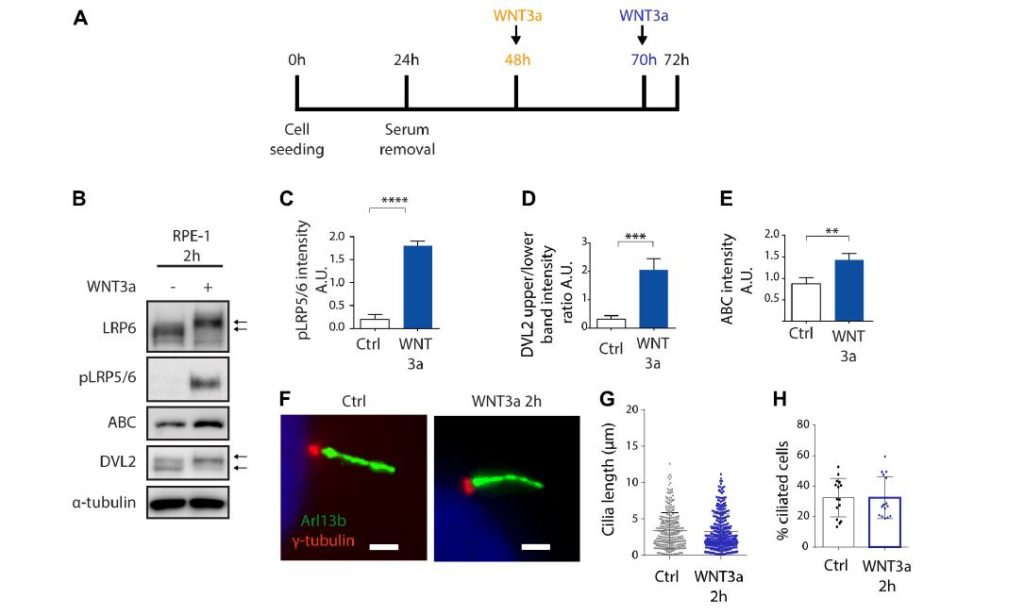
To clarify these discrepancies, we investigated possible regulation of primary cilia by the WNT/β-catenin pathway in cell lines (RPE-1, NIH3T3, and HEK293) commonly used to study ciliogenesis. We used WNT3a to activate or LGK974 to block the pathway, and examined initiation of ciliogenesis, cilium length, and percentage of ciliated cells.
We show that the treatment by WNT3a has no- or lesser inhibitory effect on cilia formation. Importantly, the inhibition of secretion of endogenous WNT ligands using LGK974 blocks WNT signaling but does not affect ciliogenesis. Finally, using knock-out cells for key WNT pathway components, namely DVL1/2/3, LRP5/6, or AXIN1/2 we show that neither activation nor deactivation of the WNT/β-catenin pathway affects the process of ciliogenesis.
These results suggest that WNT/β-catenin-mediated signaling is not generally required for efficient cilia formation. In fact, activation of the WNT/β-catenin pathway in some systems seems to moderately suppress ciliogenesis.
Front Cell Dev Biol. 2021 Feb 26;9:623753.doi: 10.3389/fcell.2021.623753. eCollection 2021.
Authors:
Ondrej Bernatik 1, Petra Paclikova 2, Anna Kotrbova 2, Vitezslav Bryja 2, Lukas Cajanek 1
- 1 Department of Histology and Embryology, Faculty of Medicine, Masaryk University, Brno, Czechia.
- 2 Section of Animal Physiology and Immunology, Department of Experimental Biology, Faculty of Science, Masaryk University, Brno, Czechia.

