Harnessing the potential of ascites to improve outcomes for ovarian cancer patients through innovative research
Extracellular vesicles in ovarian cancer and the potential of malignant ascites for ovarian cancer research
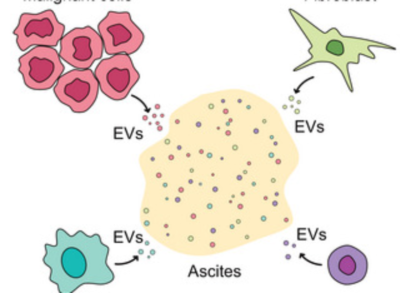
Ovarian cancer (OC) ranks among the deadliest cancers in women. Lack of symptoms, rapid metastases and common chemoresistance contribute to unfortunate fate of majority of OC patients, especially those having high-grade serous carcinoma of the ovary, fallopian tube and peritoneum (HGSC), the most common and most aggressive type of OC. Many HGSC patients have excess fluid in the peritoneum called ascites. Ascites is basically a tumor microenvironment (TME) containing various cells, proteins and extracellular vesicles (EVs).
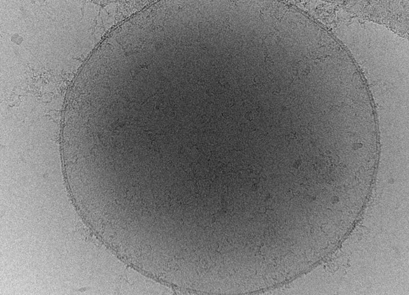
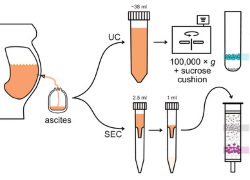
EVs play an important role in cancerogenesis and hold great promise as disease biomarkers, yet their small size and polydispersity bring various challenges to their isolation and characterization, including method-dependent enrichment of different EV subtypes as well as contaminants.
Our research projects focus on method development in the EV field, comparison of different isolation methods and especially on proteomic analyses of ascitic EVs and how these can help in unraveling the underlying molecular principles of disease progression and how can ascites serve as a new source of diagnostic/screening/prognostic EV-based biomarkers of HGSC and clinically relevant disease models, such as organoids.

We are proud members of the International Society for Extracellular Vesicles (ISEV) and co-founders of Czech Society for Extracellular Vesicles (CzeSEV).

Research
Our Team
Principal Investigator
RNDr. Vendula Hlaváčková Pospíchalová, Ph.D.

Alumni
Phd Students
Master students
- Mgr. Jana Lapinová
- Mgr. Jana Kramářová
Selected Publications
- Gregorova J, Vlachova M, Vychytilova-Faltejskova P, Dostalova A, Ruzickova T, Vecera M, Radova L, Pospichalova V, Sladecek S, Hyzdalova M, Kotaskova J, Jarosova M, Masek J, Benesova K, Jarkovsky J, Rihova L, Bezdekova R, Almasi M, Boichuk I, Stork M, Pour L, Sevcikova S. MicroRNA Profiling of Bone Marrow Plasma Extracellular Vesicles in Multiple Myeloma, Extramedullary Disease, and Plasma Cell Leukemia. Hematol Oncol. 2025 Jan;43(1):e70036. doi: 10.1002/hon.70036. PMID: 39804194; PMCID: PMC11727818.
- Drápela S, Kvokačková B, Slabáková E, Kotrbová A, Gömöryová K, Fedr R, Kurfürstová D, Eliáš M, Študent V Jr, Lenčéšová F, Ranjani GS, Pospíchalová V, Bryja V, van Weerden WM, Puhr M, Culig Z, Bouchal J, Souček K. Pre-existing cell subpopulations in primary prostate cancer tumors display surface fingerprints of docetaxel-resistant cells. Cell Oncol (Dordr). 2024 Aug 20. doi: 10.1007/s13402-024-00982-2. Epub ahead of print. PMID: 39162992.
- Welsh JA, Goberdhan DCI, O’Driscoll L, Buzas EI, Blenkiron C, Bussolati B, Cai H, Di Vizio D, Driedonks TAP, Erdbrügger U, Falcon-Perez JM, Fu QL, Hill AF, Lenassi M, Lim SK, Mahoney MG, Mohanty S, Möller A, Nieuwland R, Ochiya T, Sahoo S, Torrecilhas AC, Zheng L, Zijlstra A, Abuelreich S, Bagabas R, Bergese P, Bridges EM, Brucale M, Burger D, Carney RP, Cocucci E, Crescitelli R, Hanser E, Harris AL, Haughey NJ, Hendrix A, Ivanov AR, Jovanovic-Talisman T, Kruh-Garcia NA, Ku’ulei-Lyn Faustino V, Kyburz D, Lässer C, Lennon KM, Lötvall J, Maddox AL, Martens-Uzunova ES, Mizenko RR, Newman LA, Ridolfi A, Rohde E, Rojalin T, Rowland A, Saftics A, Sandau US, Saugstad JA, Shekari F, Swift S, Ter-Ovanesyan D, Tosar JP, Useckaite Z, Valle F, Varga Z, van der Pol E, van Herwijnen MJC, Wauben MHM, Wehman AM, Williams S, Zendrini A, Zimmerman AJ; MISEV Consortium; Théry C, Witwer KW. Minimal information for studies of extracellular vesicles (MISEV2023): From basic to advanced approaches. J Extracell Vesicles. 2024 Feb;13(2):e12404. doi: 10.1002/jev2.12404. Erratum in: J Extracell Vesicles. 2024 May;13(5):e12451. doi: 10.1002/jev2.12451. PMID: 38326288; PMCID: PMC10850029.
- Turkova K, Balvan J, Ambrozova G, Galisova A, Hyzdalova M, Tripisciano C, Cerny V, Schabussova I, Holnthoner W, Pospichalova V. A comprehensive summary of the ASEV-CzeSEV joint meeting on extracellular vesicles. Extracell Vesicles Circ Nucl Acids. 2024 Dec 21;5(4):765-784. doi: 10.20517/evcna.2024.86. PMID: 39811734; PMCID: PMC11725423.
- Vyhlídalová Kotrbová A, Gömöryová K, Mikulová A, Plešingerová H, Sladeček S, Kravec M, Hrachovinová Š, Potěšil D, Dunsmore G, Blériot C, Bied M, Kotouček J, Bednaříková M, Hausnerová J, Minář L, Crha I, Felsinger M, Zdráhal Z, Ginhoux F, Weinberger V, Bryja V, Pospíchalová V. Proteomic analysis of ascitic extracellular vesicles describes tumour microenvironment and predicts patient survival in ovarian cancer. J Extracell Vesicles. 2024 Mar;13(3):e12420. doi: 10.1002/jev2.12420. PMID: 38490958; PMCID: PMC10942866.
- Bied M, Pospíchalová V, Blériot C. Circulating extracellular vesicles as the source of diagnostic biomarkers for diseases. Clinical & Translational Discovery. 2022; 2:e145. doi.org/10.1002/ctd2.145
- Kotrbová A, Ovesná P, Gybel’ T, Radaszkiewicz T, Bednaříková M, Hausnerová J, Jandáková E, Minář L, Crha I, Weinberger V, Záveský L, Bryja V, Pospíchalová V. WNT signaling inducing activity in ascites predicts poor outcome in ovarian cancer. Theranostics. 2020 Jan 1;10(2):537-552. doi: 10.7150/thno.37423. PMID: 31903136; PMCID: PMC6929979.
- Pospichalova V, Svoboda J, Dave Z, Kotrbova A, Kaiser K, Klemova D, Ilkovics L, Hampl A, Crha I, Jandakova E, Minar L, Weinberger V, Bryja V. Simplified protocol for flow cytometry analysis of fluorescently labeled exosomes and microvesicles using dedicated flow cytometer. J Extracell Vesicles. 2015 Mar 31;4:25530. doi: 10.3402/jev.v4.25530. PMID: 25833224; PMCID: PMC4382613.
- Drápela S, Kvokačková B, Slabáková E, Kotrbová A, Gömöryová K, Fedr R, Kurfürstová D, Eliáš M, Študent V Jr, Lenčéšová F, Ranjani GS, Pospíchalová V, Bryja V, van Weerden WM, Puhr M, Culig Z, Bouchal J, Souček K. Pre-existing cell subpopulations in primary prostate cancer tumors display surface fingerprints of docetaxel-resistant cells. Cell Oncol (Dordr). 2024 Aug 20. doi: 10.1007/s13402-024-00982-2. Epub ahead of print. PMID: 39162992.
- Kotrbová A, Štěpka K, Maška M, Pálenik JJ, Ilkovics L, Klemová D, Kravec M, Hubatka F, Dave Z, Hampl A, Bryja V, Matula P, Pospíchalová V. TEM ExosomeAnalyzer: a computer-assisted software tool for quantitative evaluation of extracellular vesicles in transmission electron microscopy images. J Extracell Vesicles. 2019 Jan 21;8(1):1560808. doi: 10.1080/20013078.2018.1560808. PMID: 30719239; PMCID: PMC6346710.
- Mo L, Pospichalova V, Huang Z, Murphy SK, Payne S, Wang F, Kennedy M, Cianciolo GJ, Bryja V, Pizzo SV, Bachelder RE. Ascites Increases Expression/Function of Multidrug Resistance Proteins in Ovarian Cancer Cells. PLoS One. 2015 Jul 6;10(7):e0131579. doi: 10.1371/journal.pone.0131579. PMID: 26148191; PMCID: PMC4493087.
- Ekumi KM, Paculova H, Lenasi T, Pospichalova V, Bösken CA, Rybarikova J, Bryja V, Geyer M, Blazek D, Barboric M. Ovarian carcinoma CDK12 mutations misregulate expression of DNA repair genes via deficient formation and function of the Cdk12/CycK complex. Nucleic Acids Res. 2015 Mar 11;43(5):2575-89. doi: 10.1093/nar/gkv101. Epub 2015 Feb 20. PMID: 25712099; PMCID: PMC4357706.
- Kríz V, Pospíchalová V, Masek J, Kilander MB, Slavík J, Tanneberger K, Schulte G, Machala M, Kozubík A, Behrens J, Bryja V. β-arrestin promotes Wnt-induced low density lipoprotein receptor-related protein 6 (Lrp6) phosphorylation via increased membrane recruitment of Amer1 protein. J Biol Chem. 2014 Jan 10;289(2):1128-41. doi: 10.1074/jbc.M113.498444. Epub 2013 Nov 21. PMID: 24265322; PMCID: PMC3887180.
Collaborations
- Coming soon
Grants
- 2022-2025 National institute for cancer research (LX22NPO5102), Ministry of Education, Youth and Sports of the CR / national recovery plan
- 2024 Prognostic significance of new biomarkers identified by analysis of extracellular vesicles isolated from ascites in patients with ovarian cance (MUNI/LF-SUp/1350/2023),
Masaryk University - 2024 YIP Cancer Sectoral Meeting EMBO (1204/2024),
EMBO (European Molecular Biology Organization) / Funding for conferences and training - 2021-2024 Comprehensive genomic profiling in ovarian cancer patients treated with platinumbased chemotherapy: identification of predictive biomarkers and evaluation of the potential of precision oncology approach (NU21-03-00306),
Ministry of Health of the CR / Ministry of Health Research Programme 2020 – 2026 - 2021-2023 The potential of ascites in ovarian cancer research (MUNI/R/1225/2021), Masaryk University / Grant Agency of Masaryk University
- 2019 Význam signální dráhy Wnt v patogenezi ovariálním karcinomu (MUNI/E/0532/2019), Masaryk University / Grant Agency of Masaryk University
- 2017-2019 Funkční a biochemická analýza extracelulárních váčků z ascitů pacientek s karcinomem vaječníku (GJ17-11776Y), Czech Science Foundation / Junior projects
- 2018 TEM ExosomeAnalyzer: Nový software pro kvantitativní charakterizaci extracelulárních vesikulů pomocí transmisní elektronové mikroskopie (MUNI/E/0876/2018), Masaryk University / Grant Agency of Masaryk University
- 2014-2016 Automatizovaná analýza elektronmikroskopických snímků pro použití v biologii a medicíně (MUNI/M/1050/2013), Masaryk University / Grant Agency of Masaryk University
- 2012-2015 Zaměstnáním čerstvých absolventů doktorského studia k vědecké excelenci (CZ.1.07/2.3.00/30.0009), Ministry of Education, Youth and Sports of the CR / Operational Programme Education for Competitiveness
Media
Conferences and Poster Presentations
- Coming soon


